A) joints.
B) bursae.
C) sutures.
D) cartilage.
F) None of the above
Correct Answer

verified
Correct Answer
verified
Multiple Choice
What is the central-ray angulation for the SMV projection?
A) 0 degrees
B) 5 degrees caudad
C) 5 degrees cephalad
D) 5 to 7 degrees cephalad
F) A) and D)
Correct Answer

verified
Correct Answer
verified
Multiple Choice
For the tangential projection of the zygomatic arch,the central ray is directed perpendicular to the _____ line.
A) infraorbitomeatal
B) mentomeatal
C) orbitomeatal
D) acanthiomeatal
F) A) and B)
Correct Answer

verified
Correct Answer
verified
Multiple Choice
The bones of the cranium are joined together by fibrous joints called:
A) diploë.
B) sulci.
C) sutures.
D) cartilage.
F) All of the above
Correct Answer

verified
Correct Answer
verified
Multiple Choice
The letter A in the image below of the paranasal sinuses labels the _____ sinuses. 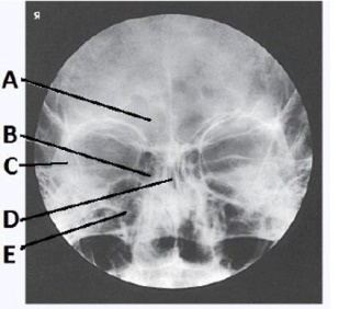
A) ethmoid
B) sphenoid
C) maxillary
D) frontal
F) A) and B)
Correct Answer

verified
Correct Answer
verified
Multiple Choice
How was the central ray directed to obtain the image below,used to evaluate the cranium? 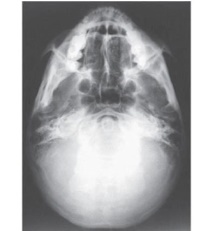
A) Perpendicular to IOML
B) 15 degrees caudad
C) 25 degrees cephalad
D) 37 degrees caudad
F) B) and C)
Correct Answer

verified
Correct Answer
verified
Multiple Choice
The part of the sphenoid bone identified in the figure below is the: 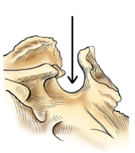
A) clivus.
B) foramen magnum.
C) sella turcica.
D) dorsum sellae.
F) A) and C)
Correct Answer

verified
Correct Answer
verified
Multiple Choice
Which two are clearly demonstrated within the foramen magnum during an AP axial (Towne) projection of the skull? (Select all that apply.)
A) Dorsum sellae
B) Posterior clinoids
C) Sella turcica
D) Anterior clinoids
F) B) and C)
Correct Answer

verified
Correct Answer
verified
Multiple Choice
Where is the image receptor centered for the parietoacanthial (Waters method) projection of the sinuses?
A) Nasion
B) Inion
C) Glabella
D) Acanthion
F) B) and C)
Correct Answer

verified
Correct Answer
verified
Multiple Choice
Which of the following is located in the internal ear?
A) Concha
B) Auditory tube
C) Tympanic membrane
D) Semicircular canals
F) B) and C)
Correct Answer

verified
Correct Answer
verified
Multiple Choice
What projection (method) used to evaluate the cranium is demonstrated in the image below? 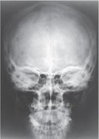
A) Parietoacanthial (Waters)
B) PA axial (Caldwell)
C) AP axial (Towne)
D) PA
F) B) and D)
Correct Answer

verified
Correct Answer
verified
Multiple Choice
What structure is labeled as letter B in the image below,used to evaluate the cranium? 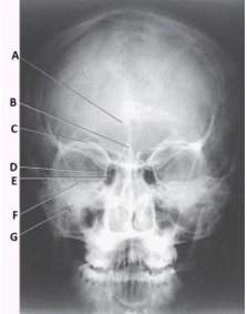
A) Acanthion
B) Crista galli
C) Cribriform plate
D) Petrous ridge
F) A) and D)
Correct Answer

verified
Correct Answer
verified
Multiple Choice
All are clearly demonstrated on an SMV projection of the cranial base,except:
A) carotid canals.
B) sphenoid sinuses.
C) mastoid process.
D) frontal sinuses.
F) C) and D)
Correct Answer

verified
Correct Answer
verified
Multiple Choice
The letter D in the image below of the paranasal sinuses labels the _____ sinuses. 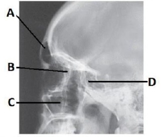
A) frontal
B) ethmoid
C) sphenoid
D) maxillary
F) B) and C)
Correct Answer

verified
Correct Answer
verified
Multiple Choice
The midsagittal plane of the head is rotated how many degrees for the axiolateral oblique projection of the TMJ?
A) 0 degrees toward the IR
B) 15 degrees toward the IR
C) 0 degrees away from the IR
D) 15 degrees away from the IR
F) B) and C)
Correct Answer

verified
Correct Answer
verified
Multiple Choice
The bone identified on the skull below is the: 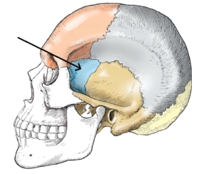
A) temporal.
B) parietal.
C) zygoma.
D) sphenoid.
F) B) and D)
Correct Answer

verified
Correct Answer
verified
Multiple Choice
Often a patient cannot be turned into the prone position for a PA axial projection of the skull (Caldwell method) .What central-ray angle would be used if the AP axial projection is used instead?
A) 10 degrees caudad
B) 15 degrees cephalad
C) 10 to 15 degrees caudad
D) 10 to 15 degrees cephalad
F) A) and B)
Correct Answer

verified
Correct Answer
verified
Multiple Choice
Radiographic demonstration of the cranial base is performed by which method?
A) Haas
B) Rhese
C) Towne
D) Schüller
F) C) and D)
Correct Answer

verified
Correct Answer
verified
Multiple Choice
Which reference line is positioned horizontal to ensure proper extension of the head during a lateral projection of the sinuses?
A) AML
B) OML
C) IOML
D) MML
F) B) and D)
Correct Answer

verified
Correct Answer
verified
Multiple Choice
Where are the petrous ridges seen on an image of a parietoacanthial (Waters method) projection of the paranasal sinuses?
A) In the middle of the maxillary sinuses
B) Superior to the maxillary sinuses
C) Inferior to the floor of the maxillary sinuses
D) In the lower two thirds of the maxillary sinuses
F) All of the above
Correct Answer

verified
Correct Answer
verified
Showing 101 - 120 of 185
Related Exams